INTRODUCTION:
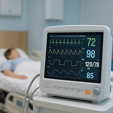
A patient monitor is a vital medical device used in hospitals and healthcare settings to continuously observe a patient’s physiological conditions. It is the most important equipment for critical tools. It helps healthcare professionals detect changes in a patient’s health early and respond quickly. Patient monitors are commonly used in intensive care units (ICUs), operation theaters, emergency rooms, and general wards.
WHAT IS A PATIENT MONITOR?
A patient monitor is an electronic device that measures, displays, and records important vital signs of a patient. These vital signs indicate how well the body’s essential systems are functioning. The monitor provides real-time data, allowing medical staff to assess the patient’s condition accurately.
VITALS MEASURED BY A PATIENT MONITOR
1) BLOOD PRESSURE:
Blood pressure measures the force of blood against the walls of the arteries. It is usually expressed as systolic pressure over diastolic pressure.
120 mmHg→systolic
80 mmHg→diastolic
Blood pressure can be measured by using two methods:
➢ NON-INVASIVE BLOOD PRESSURE(NIBP) This method measures blood pressure externally, usually with an inflatable cuff placed around the arm.
➢ INVASIVE BLOOD PRESSURE(IBP) This method measures blood pressure internally using an arterial catheter. It is most commonly used in operation theaters and intensive care units for continuous and accurate monitoring.
2) HEART RATE:
Heart rate represents the number of heart beats per minute. It reflects how effectively the heart is pumping blood. Abnormal heart rates may indicate conditions such as arrhythmia, stress, or cardiac disease.
• 60–100 bpm: Normal resting heart rate for adults
• 50–60 bpm: Normal resting heart rate in well-trained athletes
• 60–80 bpm: Typical resting range for a healthy adult
• 100–180 bpm: May occur during physical activity or exercise A resting heart rate of around 72 bpm is often considered ideal for healthy adults.
3) OXYGEN SATURATION (Sp𝑶𝟐)
SpO₂ measures the percentage of oxygen carried by hemoglobin in the blood. It reflects respiratory efficiency and oxygen delivery to tissues.
• Normal range: 95–100%
• Below 90%: May indicate hypoxemia and requires urgent medical attention
In a patient monitor, oxygen saturation is measured using a pulse oximeter probe, usually attached to a finger, toe, or earlobe. The probe works by emitting red and infrared light, which passes through the tissue to detect oxygenated and deoxygenated hemoglobin, allowing the monitor to calculate SpO₂ accurately.
4) BODY TEMPERATURE:
Body temperature indicates the body’s thermal balance and metabolic state.
• Normal range: 36.1–37.2°C (97–99°F)
• Fever: Above 37.5°C (99.5°F)
• Hypothermia: Below 35°C (95°F)
In a patient monitor, body temperature is measured using a temperature probe that contains a thermistor. The thermistor converts heat energy from the body into an electrical signal, which the monitor interprets to display the patient’s temperature accurately.
5) ELECTROCARDIOGRAM:
An ECG measures the electrical activity of the heart. It helps detect heart rhythm abnormalities, conduction problems, and signs of heart disease.
• The patient monitor records electrical signals from the heart using electrodes placed on the chest, arms, and legs.
• The signals are displayed as waveforms (P wave, QRS complex, T wave), which indicate the timing of atrial and ventricular contractions.
• Continuous ECG monitoring allows healthcare professionals to detect arrhythmias, ischemia, or other cardiac events early, especially in critically ill patients.
Some patient monitors have 3 leads for basic monitoring, while others have 5 leads for advanced monitoring.
• 3-lead ECG: Measures basic heart rhythm and is commonly used for general monitoring in wards or during transport. It provides limited but sufficient information about the heart’s electrical activity.
• 5-lead ECG: Provides more detailed information and allows monitoring of both heart rhythm and more accurate detection of ischemic changes. This is often used in ICUs, operation theaters, and critical care.
In an ECG, aVR, aVL, and aVF are augmented limb leads that provide different views of the heart’s electrical activity.
• aVR (augmented Vector Right): Measures the electrical activity from the heart toward the right arm. It mainly helps identify abnormal conduction and the overall direction of electrical impulses.
• aVL (augmented Vector Left): Measures the electrical activity from the heart toward the left arm. It provides a view of the lateral wall of the left ventricle.
• aVF (augmented Vector Foot): Measures the electrical activity from the heart toward the left leg (foot). It provides a view of the inferior wall of the heart.